Magnetic Resonance - Technology Information Portal Welcome to MRI Technology
Info
  Sheets

Out-
      side


 'Fast Spoiled Gradient Echo' 
SEARCH FOR    
 
  2 3 5 A B C D E F G H I J K L M N O P Q R S T U V W X Y Z
Result : Searchterm 'Fast Spoiled Gradient Echo' found in 1 term [] and 0 definition [], (+ 11 Boolean[] results
1 - 5 (of 12)     next
Result Pages : [1]  [2 3]
MRI Resources 
Bioinformatics - Health - MRI Centers - Jobs - Shoulder MRI - Lung Imaging
 
Fast Spoiled Gradient EchoInfoSheet: - Sequences - 
Intro, 
Overview, 
Types of, 
etc.
 
(FSPGR) A sequence similar to TurboFLASH or Turbo Field Echo.

See also Spoiled Gradient Echo Sequence and Ultrafast Gradient Echo Sequence.
spacer
 
• Share the entry 'Fast Spoiled Gradient Echo':  Facebook  Twitter  LinkedIn  
 
Further Reading:
  News & More:
3-D VOLUMETRIC IMAGING FOR STEREOTACTIC LESIONAL AND DEEP BRAIN STIMULATION SURGERY
MRI Resources 
Mobile MRI - Liver Imaging - MRI Centers - Patient Information - Breast MRI - Blood Flow Imaging
 
Virgo™InfoSheet: - Devices -
Intro, 
Types of Magnets, 
Overview, 
etc.MRI Resource Directory:
 - Devices -
 
www.canamglobal.com/mri/virgomri.html From Millennium Technology Inc. This open C-shaped MRI system eases patient comfort and technologist maneuverability. This low cost scanner is build for a wide range of applications. The Virgo™ patient table is detachable and moves on easy rolling castors. Able to accommodate patient weights up to 160 kg, the tabletop has a range of motion of 30 cm in the lateral direction and 90cm in the longitudinal direction. Images generated with this scanner can only be viewed (without data loss) on Millennium's proprietary viewing software.
Device Information and Specification
CLINICAL APPLICATION
Whole body
CONFIGURATION
C shaped
SYNCHRONIZATION
Standard cardiac gating, ECG/peripheral, respiratory gating
PULSE SEQUENCES
2D Spin Echo (single and multi-echo), 2D Inversion Recovery, 2D Sequential and 3D Volume Gradient Echo, 2D and 3D Spoiled Gradient Echo
IMAGING MODES
Localizer, single slice, multislice, volume, fast, POMP, multi slab, cine, slice and frequency zip, extended dynamic range, tailored RF
TR
steps of 1 msec
TE
steps of 1 msec
SINGLE/MULTI SLICE
Simultaneous scan and reconstruction;; 100 images/second reconstruction
400 mm
2D : 2 mm; 3D : 0.5 mm
MEASURING MATRIX
512x512
PIXEL INTENSITY
256 gray levels
MAGNET TYPE
Permanent
BORE DIAMETER
or Vertical Gap
44 cm
STRENGTH
15 mT/m
5-GAUSS FRINGE FIELD
3 m/3 m
Passive
spacer
MRI Resources 
Nerve Stimulator - MRI Physics - PACS - Portals - Supplies - Safety Products
 
Gradient Echo SequenceForum -
related threadsInfoSheet: - Sequences - 
Intro, 
Overview, 
Types of, 
etc.
 
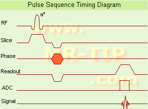
Gradient Echo Sequence Timing Diagram (GRE - sequence) A gradient echo is generated by using a pair of bipolar gradient pulses. In the pulse sequence timing diagram, the basic gradient echo sequence is illustrated. There is no refocusing 180° pulse and the data are sampled during a gradient echo, which is achieved by dephasing the spins with a negatively pulsed gradient before they are rephased by an opposite gradient with opposite polarity to generate the echo.
See also the Pulse Sequence Timing Diagram. There you will find a description of the components.
The excitation pulse is termed the alpha pulse α. It tilts the magnetization by a flip angle α, which is typically between 0° and 90°. With a small flip angle there is a reduction in the value of transverse magnetization that will affect subsequent RF pulses. The flip angle can also be slowly increased during data acquisition (variable flip angle: tilt optimized nonsaturation excitation). The data are not acquired in a steady state, where z-magnetization recovery and destruction by ad-pulses are balanced. However, the z-magnetization is used up by tilting a little more of the remaining z-magnetization into the xy-plane for each acquired imaging line.
Gradient echo imaging is typically accomplished by examining the FID, whereas the read gradient is turned on for localization of the signal in the readout direction. T2* is the characteristic decay time constant associated with the FID. The contrast and signal generated by a gradient echo depend on the size of the longitudinal magnetization and the flip angle. When α = 90° the sequence is identical to the so-called partial saturation or saturation recovery pulse sequence. In standard GRE imaging, this basic pulse sequence is repeated as many times as image lines have to be acquired. Additional gradients or radio frequency pulses are introduced with the aim to spoil to refocus the xy-magnetization at the moment when the spin system is subject to the next α pulse.
As a result of the short repetition time, the z-magnetization cannot fully recover and after a few initial α pulses there is an equilibrium established between z-magnetization recovery and z-magnetization reduction due to the α pulses.
Gradient echoes have a lower SAR, are more sensitive to field inhomogeneities and have a reduced crosstalk, so that a small or no slice gap can be used. In or out of phase imaging depending on the selected TE (and field strength of the magnet) is possible. As the flip angle is decreased, T1 weighting can be maintained by reducing the TR. T2* weighting can be minimized by keeping the TE as short as possible, but pure T2 weighting is not possible. By using a reduced flip angle, some of the magnetization value remains longitudinal (less time needed to achieve full recovery) and for a certain T1 and TR, there exist one flip angle that will give the most signal, known as the "Ernst angle".
Contrast values:
PD weighted: Small flip angle (no T1), long TR (no T1) and short TE (no T2*)
T1 weighted: Large flip angle (70°), short TR (less than 50ms) and short TE
T2* weighted: Small flip angle, some longer TR (100 ms) and long TE (20 ms)

Classification of GRE sequences can be made into four categories:
See also Gradient Recalled Echo Sequence, Spoiled Gradient Echo Sequence, Refocused Gradient Echo Sequence, Ultrafast Gradient Echo Sequence.
 
Images, Movies, Sliders:
 MRI Liver In Phase  Open this link in a new window
    
 MRI Liver Out Of Phase  Open this link in a new window
    
 MVP Parasternal  Open this link in a new window
 Breast MRI Images T1 Pre - Post Contrast  Open this link in a new window
 Circle of Willis, Time of Flight, MIP  Open this link in a new window
    
SlidersSliders Overview

 
spacer

• View the DATABASE results for 'Gradient Echo Sequence' (70).Open this link in a new window

 
Further Reading:
  Basics:
Enhanced Fast GRadient Echo 3-Dimensional (efgre3D) or THRIVE
   by www.mri.tju.edu    
  News & More:
MRI evaluation of fatty liver in day to day practice: Quantitative and qualitative methods
Wednesday, 3 September 2014   by www.sciencedirect.com    
T1rho-prepared balanced gradient echo for rapid 3D T1rho MRI
Monday, 1 September 2008   by www.ncbi.nlm.nih.gov    
MRI Resources 
IR - Distributors - Manufacturers - Nerve Stimulator - Safety pool - Mass Spectrometry
 
Blood Oxygenation Level Dependent ContrastInfoSheet: - Sequences - 
Intro, 
Overview, 
Types of, 
etc.MRI Resource Directory:
 - Functional MRI -
 
(BOLD) In MRI the changes in blood oxygenation level are visible. Oxyhaemoglobin (the principal haemoglobin in arterial blood) has no substantial magnetic properties, but deoxyhaemoglobin (present in the draining veins after the oxygen has been unloaded in the tissues) is strongly paramagnetic. It can thus serve as an intrinsic paramagnetic contrast agent in appropriately performed brain MRI. The concentration and relaxation properties of deoxyhaemoglobin make it a susceptibility , e.g. T2 relaxation effective contrast agent with little effect on T1 relaxation.
During activation of the brain, the oxygen consumption of the local tissue increase by approximately 5% with that the oxygen tension will decrease. As a consequence, after a short period of time vasodilatation occurs, resulting in a local increase of blood volume and flow by 20 - 40%. The incommensurate change in local blood flow and oxygen extraction increases the local oxygen level.
By using T2 weighted gradient echo EPI sequences, which are highly susceptibility sensitive and fast enough to capture the three-dimensional nature of activated brain areas will show an increase in signal intensity as oxyhaemoglobin is diamagnetic and deoxyhaemoglobin is paramagnetic. Other MR pulse sequences, such as spoiled gradient echo pulse sequences are also used.
As the effects are subtle and of the order of 2% in 1.5 T MR imaging, sophisticated methodology, paradigms and data analysis techniques have to be used to consistently demonstrate the effect.
As the BOLD effect is due to the deoxygenated blood in the draining veins, the spatial localization of the region where there is increased blood flow resulting in decreased oxygen extraction is not as precisely defined as the morphological features in MRI. Rather there is a physiological blurring, and is estimated that the linear dimensions of the physiological spatial resolution of the BOLD phenomenon are around 3 mm at best.
spacer

• View the DATABASE results for 'Blood Oxygenation Level Dependent Contrast' (6).Open this link in a new window

 
Further Reading:
  Basics:
IMAGE CONTRAST IN MRI(.pdf)
   by www.assaftal.com    
Vascular Filters of Functional MRI: Spatial Localization Using BOLD and CBV Contrast
  News & More:
A mechanistic computational framework to investigate the hemodynamic fingerprint of the blood oxygenation level-dependent signal
Tuesday, 29 August 2023   by analyticalsciencejournals.onlinelibrary.wiley.com    
The utility of texture analysis of kidney MRI for evaluating renal dysfunction with multiclass classification model
Tuesday, 30 August 2022   by www.nature.com    
MRI Technique Used to Identify Future Risk of Binge Drinking
Monday, 6 January 2020   by www.diagnosticimaging.com    
Gold Acupuncture Needle MRI Pain Discovery
Friday, 3 January 2014   by www.healthcmi.com    
MRI method for measuring MS progression validated
Thursday, 19 December 2013   by www.eurekalert.org    
MRI Resources 
Service and Support - Colonography - MRI Training Courses - Mass Spectrometry - Process Analysis - Claustrophobia
 
Fast Low Angle ShotInfoSheet: - Sequences - 
Intro, 
Overview, 
Types of, 
etc.MRI Resource Directory:
 - Sequences -
 
(FLASH) A fast sequence producing signals called gradient echo with low flip angles. FLASH sequences are modifications, which incorporate or remove the effects of transverse coherence respectively.
FLASH uses a semi-random spoiler gradient after each echo to spoil the steady state (to destroy any remaining transverse magnetization) by causing a spatially dependent phase shift. The transverse steady state is spoiled but the longitudinal steady state depends on the T1 values and the flip angle. Extremely short TR times are possible, as a result the sequence provides a mechanism for gaining extremely high T1 contrast by imaging with TR times as brief as 20 to 30 msec while retaining reasonable signal levels. It is important to keep the TE as short as possible to suppress susceptibility artifacts.
The T1 contrast depends on the TR as well as on flip angle, with short TE.
Small flip angles and short TR results in proton density, and long TR in T2* weighting.
With large flip angles and short TR result T1 weighted images.

TR and flip angle adjustment:

TR 3000 ms, Flip Angle 90°
TR 1500 ms, Flip Angle 45°
TR 700 ms, Flip Angle 25°
TR 125 ms, Flip Angle 10°

The apparent ability to trade TR against flip angle for purposes of contrast and the variation in SNR as the scan time (TR) is reduced.

See also Gradient Echo Sequence.
 
Images, Movies, Sliders:
 Fetus (Brain) and Dermoid in Mother  Open this link in a new window
      

Courtesy of  Robert R. Edelman

 
spacer

• View the DATABASE results for 'Fast Low Angle Shot' (5).Open this link in a new window

 
Further Reading:
  News & More:
Motion Compensation in MR Imaging
   by ccn.ucla.edu    
Turbo-FLASH Based Arterial Spin Labeled Perfusion MRI at 7 T
Thursday, 20 June 2013   by www.plosone.org    
Usefulness of MR Imaging for Diseases of the Small Intestine: Comparison with CT
2000   by www.ncbi.nlm.nih.gov    
MRI Resources 
Examinations - Claustrophobia - MRI Technician and Technologist Career - MRI Accidents - IR - Jobs pool
 
     1 - 5 (of 12)     next
Result Pages : [1]  [2 3]
 Random Page
 
Share This Page
FacebookTwitterLinkedIn

MR-TIP    
Community   
User
Pass
Forgot your UserID/Password ?    


New acceleration techniques will :
reduce scan times 
cause artifacts 
increase expenses 
be useful if you have a lot of experience 
doesn't do much 
never heard of 

Look
      Ups


MR-TIP.com uses cookies! By browsing MR-TIP.com, you agree to our use of cookies.

Magnetic Resonance - Technology Information Portal
Member of SoftWays' Medical Imaging Group - MR-TIP • Radiology-TIP • Medical-Ultrasound-Imaging • 
Copyright © 2003 - 2024 SoftWays. All rights reserved. [ 18 April 2024]
Terms of Use | Privacy Policy | Advertising
 [last update: 2024-02-26 03:41:00]