Magnetic Resonance - Technology Information Portal Welcome to MRI Technology
Info
  Sheets

Out-
      side


 'Dual Echo Sequence' 
SEARCH FOR    
 
  2 3 5 A B C D E F G H I J K L M N O P Q R S T U V W X Y Z
Result : Searchterm 'Dual Echo Sequence' found in 1 term [] and 2 definitions [], (+ 6 Boolean[] results
1 - 5 (of 9)     next
Result Pages : [1]  [2]
MRI Resources 
Directories - - IR - Pediatric and Fetal MRI - MRI Centers - Artifacts
 
Dual Echo SequenceInfoSheet: - Sequences - 
Intro, 
Overview, 
Types of, 
etc.
 
(DE - dual / double echo) Dual echo sequences include images with different weightings and / or echo times and are used to obtain both, proton density and T2 weighted images or in phase and out of phase gradient echo images, simultaneously without increasing the measurement time.
 
Images, Movies, Sliders:
 MRI Liver In Phase  Open this link in a new window
 MRI Liver Out Of Phase  Open this link in a new window
 
spacer
 
• Share the entry 'Dual Echo Sequence':  Facebook  Twitter  LinkedIn  
 
• Related Searches:
    • Opposed Phase Image
    • Black Boundary Artifact
    • Chemical Shift Artifact
    • Double Inversion Recovery T1 Measurement
    • Gradient Echo
 
Further Reading:
  Basics:
BASIC PRINCIPLES OF MR IMAGING
   by spinwarp.ucsd.edu    
MRI evaluation of fatty liver in day to day practice: Quantitative and qualitative methods
Wednesday, 3 September 2014   by www.sciencedirect.com    
  News & More:
Iron overload: accuracy of in-phase and out-of-phase MRI as a quick method to evaluate liver iron load in haematological malignancies and chronic liver disease
Friday, 1 June 2012   by www.ncbi.nlm.nih.gov    
MRI Resources 
Sequences - Pathology - Cardiovascular Imaging - Pacemaker - Process Analysis - Libraries
 
Dual Echo Steady StateInfoSheet: - Sequences - 
Intro, 
Overview, 
Types of, 
etc.
 
(DESS) This sequence was originally known as FADE. It combines both the gradient echoes acquired in FISP and PSIF sequences in separate acquisition periods during a single interpulse interval. Phase encoding gradients are balanced to maintain the transverse steady state signals. The frequency encoding gradient is left on for the period of both the echoes, and is incompletely balanced to avoid dark banding artifacts otherwise associated with long TR fully balanced steady state sequences. The contrast of DESS is quite unique, true T2 or T1 contrast weighting is not possible. There is a strong fluid signal but fat is bright and other soft tissues appear similar to the short TR FISP image.
Used for, e.g. the joints, cartilage and the prostate.

See Steady State Free Precession and Dual Echo Sequence.
spacer

• View the DATABASE results for 'Dual Echo Steady State' (2).Open this link in a new window

MRI Resources 
Implant and Prosthesis pool - PACS - Breast Implant - Pediatric and Fetal MRI - Mobile MRI - Movies
 
Fast Spin EchoForum -
related threadsInfoSheet: - Sequences - 
Intro, 
Overview, 
Types of, 
etc.
 
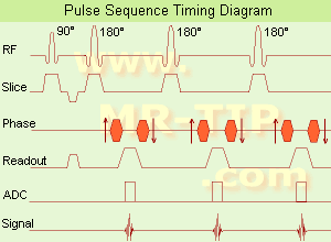
Fast Spin Echo Diagram (FSE) In the pulse sequence timing diagram, a fast spin echo sequence with an echo train length of 3 is illustrated. This sequence is characterized by a series of rapidly applied 180° rephasing pulses and multiple echoes, changing the phase encoding gradient for each echo.
The echo time TE may vary from echo to echo in the echo train. The echoes in the center of the K-space (in the case of linear k-space acquisition) mainly produce the type of image contrast, whereas the periphery of K-space determines the spatial resolution. For example, in the middle of K-space the late echoes of T2 weighted images are encoded. T1 or PD contrast is produced from the early echoes.
The benefit of this technique is that the scan duration with, e.g. a turbo spin echo turbo factor / echo train length of 9, is one ninth of the time. In T1 weighted and proton density weighted sequences, there is a limit to how large the ETL can be (e.g. a usual ETL for T1 weighted images is between 3 and 7). The use of large echo train lengths with short TE results in blurring and loss of contrast. For this reason, T2 weighted imaging profits most from this technique.
In T2 weighted FSE images, both water and fat are hyperintense. This is because the succession of 180° RF pulses reduces the spin spin interactions in fat and increases its T2 decay time. Fast spin echo (FSE) sequences have replaced conventional T2 weighted spin echo sequences for most clinical applications. Fast spin echo allows reduced acquisition times and enables T2 weighted breath hold imaging, e.g. for applications in the upper abdomen.
In case of the acquisition of 2 echoes this type of a sequence is named double fast spin echo / dual echo sequence, the first echo is usually density and the second echo is T2 weighted image. Fast spin echo images are more T2 weighted, which makes it difficult to obtain true proton density weighted images. For dual echo imaging with density weighting, the TR should be kept between 2000 - 2400 msec with a short ETL (e.g., 4).
Other terms for this technique are:
Turbo Spin Echo
Rapid Imaging Spin Echo,
Rapid Spin Echo,
Rapid Acquisition Spin Echo,
Rapid Acquisition with Refocused Echoes
 
Images, Movies, Sliders:
 Lumbar Spine T2 FSE Sagittal  Open this link in a new window
    

Courtesy of  Robert R. Edelman
 MRI - Anatomic Imaging of the Foot  Open this link in a new window
    
SlidersSliders Overview

 Lumbar Spine T2 FSE Axial  Open this link in a new window
    

Courtesy of  Robert R. Edelman
 
spacer

• View the DATABASE results for 'Fast Spin Echo' (31).Open this link in a new window

 
Further Reading:
  Basics:
MYELIN-SELECTIVE MRI: PULSE SEQUENCE DESIGN AND OPTIMIZATION
   by www.imaging.robarts.ca    
Advances in Magnetic Resonance Neuroimaging
Friday, 27 February 2009   by www.ncbi.nlm.nih.gov    
  News & More:
New MR sequence helps radiologists more accurately evaluate abnormalities of the uterus and ovaries
Thursday, 23 April 2009   by www.eurekalert.org    
Spin echoes, CPMG and T2 relaxation - Introductory NMR & MRI from Magritek
2013   by www.azom.com    
MRI Resources 
MRI Training Courses - MRI Physics - Mobile MRI - Chemistry - Pacemaker - MRI Centers
 
O-SCAN™InfoSheet: - Devices -
Intro, 
Types of Magnets, 
Overview, 
etc.
 
www.fonar.com/standup.htm www.fonar.com/standup.htm O-scan is manufactured and distributed by Esaote SpA
O-scan is a compact, dedicated extremity MRI system designed for easy installation and high throughput. The complete system fits in a 9' x 10' room, doesn't need for RF or magnetic shielding and it plugs in the wall. The 0.31T permanent magnet along with dual phased array RF coils, and advanced imaging protocols provide outstanding image quality and fast 25 minute complete examinations.
Esaote North America is the exclusive distributor of the O-scan system in the USA.
Device Information and Specification
CLINICAL APPLICATION
Dedicated Extremity
CONFIGURATION
Closed
Dual phased array knee, hand, foot//ankle/elbow
PULSE SEQUENCES
SE, HSE, HFE, GE, 2dGE, ME, IR, STIR, Stir T2, GESTIR, TSE, TME, FSE STIR, FSE (T1, T2), X-Bone, Turbo 3DT1, 3D SHARC, 3D SST1, 3D SST2
IMAGING MODES
2D, 3D multi-plane, half echo, half scan, real time
TR
10 - 10,000 msec.
TE
6 - 220 msec.
SINGLE SLICE
0.1 sec.
MULTI SLICE
0.1 sec.
14 cm
2D: 2mm - 10 mm, 3D: 0.6 - 10 mm
MEASURING MATRIX
512 x 512 max.
PIXEL INTENSITY
4,096 grey levels
MAGNET TYPE
Permanent - NdFeB
MAGNET WEIGHT
2,733 lbs
POWER REQUIREMENTS
100/110/200/220/230/240
STRENGTH
20 mT/m
5 GAUSS FRINGE FIELD, radial/axial
67 cm / 75 cm
passive
spacer
MRI Resources 
Databases - Developers - Shielding - MR Myelography - Guidance - Journals
 
Half Fourier Acquisition Single Shot Turbo Spin EchoInfoSheet: - Sequences - 
Intro, 
Overview, 
Types of, 
etc.MRI Resource Directory:
 - Sequences -
 
(HASTE) A pulse sequence with data acquisition after an initial preparation pulse for contrast enhancement with the use of a very long echo train (Single shot TSE), whereat each echo is individually phase encoded. This technique is a heavily T2 weighted, high speed sequence with partial Fourier technique, a great sensitivity for fluid detection and a fast acquisition time of about 1 sec per slice. This advantage makes it possible for using breath-hold with excellent motionless MRI, e.g. used for liver and lung imaging.

See also Segmented HASTE.
 
Images, Movies, Sliders:
 Anatomic Imaging of the Lungs  Open this link in a new window
      

Courtesy of  Robert R. Edelman
 Fetus (Brain) and Dermoid in Mother  Open this link in a new window
      

Courtesy of  Robert R. Edelman

 
spacer

• View the DATABASE results for 'Half Fourier Acquisition Single Shot Turbo Spin Echo' (5).Open this link in a new window

 
Further Reading:
  News & More:
EVALUATION OF HUMAN STROKE BY MR IMAGING
2000
The cerebello-pontine angle, ACNR • VOLUME 2 NUMBER 3, Page 16
2002
MRI Resources 
Implant and Prosthesis pool - Quality Advice - RIS - Lung Imaging - Cochlear Implant - Colonography
 
     1 - 5 (of 9)     next
Result Pages : [1]  [2]
 Random Page
 
Share This Page
FacebookTwitterLinkedIn

MR-TIP    
Community   
User
Pass
Forgot your UserID/Password ?    


Personalized protocols (age, gender, body habitus, etc.) lead to :
more automated planning 
improved patient comfort 
better diagnostics 
optimized image quality 
nothing 

Look
      Ups


MR-TIP.com uses cookies! By browsing MR-TIP.com, you agree to our use of cookies.

Magnetic Resonance - Technology Information Portal
Member of SoftWays' Medical Imaging Group - MR-TIP • Radiology-TIP • Medical-Ultrasound-Imaging • 
Copyright © 2003 - 2024 SoftWays. All rights reserved. [ 18 April 2024]
Terms of Use | Privacy Policy | Advertising
 [last update: 2024-02-26 03:41:00]