Magnetic Resonance - Technology Information Portal Welcome to MRI Technology
Info
  Sheets

Out-
      side


 'Diffusion Time' 
SEARCH FOR    
 
  2 3 5 A B C D E F G H I J K L M N O P Q R S T U V W X Y Z
Result : Searchterm 'Diffusion Time' found in 1 term [] and 1 definition [], (+ 17 Boolean[] results
1 - 5 (of 19)     next
Result Pages : [1]  [2 3 4]
Searchterm 'Diffusion Time' was also found in the following services: 
spacer
News  (2)  Resources  (1)  
 
Diffusion Time
 
Images obtained by a diffusion weighted sequence (e.g. the Stejskal-Tanner sequence) reflect the diffusion as an attenuation of signal intensity. The diffusion time is the time elapsed between the leading edges of the diffusion gradient lobes of this sequence.
spacer
 
• Share the entry 'Diffusion Time':  Facebook  Twitter  LinkedIn  

• View the NEWS results for 'Diffusion Time' (1).Open this link in a new window.
 
Further Reading:
  Basics:
Diffusion-Weighted MRI in the Body: Applications and Challenges in Oncology
Friday, 1 June 2007   by www.ajronline.org    
MRI Resources 
Homepages - Process Analysis - Directories - Portals - Nerve Stimulator - Coils
 
Diffusion Weighted SequenceInfoSheet: - Sequences - 
Intro, 
Overview, 
Types of, 
etc.MRI Resource Directory:
 - Diffusion Weighted Imaging -
 
Diffusion weighted imaging can be performed similar to the phase contrast angiography sequence. The gradients must be increased in amplitude to depict the much slower motions of molecular diffusion in the body.
While a T1 weighted MRI pulse sequence is diffusion sensitive, a quantitative diffusion pulse sequence was introduced by Steijskal and Tanner. Its characteristic features are two strong symmetrical gradient lobes placed on either side of the 180° refocusing pulse in a spin echo sequence. These symmetrical gradient lobes have the sole purpose of enhancing dephasing of spins, thereby accelerating intravoxel incoherent motion (IVIM) signal loss.
Dephasing is proportional to the square of the time (diffusion time) during which the gradients are switched on and the strength of the applied gradient field. Therefore, the use of high field gradient systems with faster and more sensitive sequences, make diffusion weighting more feasible.
Areas in which the protons diffuse rapidly (swollen cells in early stroke, less restriction to diffusion) will show an increased signal when the echo is measured relative to areas in which diffusion is restricted. For increased accuracy of diffusion measurement and image enhancement, useful motion correction techniques such as navigator echo and other methods should be used. In addition to this, applying the b-value calculated by the strength and duration of motion probing gradients with a high rate of accuracy is very important.

See also Apparent Diffusion Coefficient, ADC Map, Lattice Index Map.
spacer

• View the DATABASE results for 'Diffusion Weighted Sequence' (6).Open this link in a new window

 
Further Reading:
  Basics:
Diffusion-Weighted Imaging
   by spinwarp.ucsd.edu    
A Comparison of Methods for High-Spatial-Resolution Diffusion-weighted Imaging in Breast MRI
Tuesday, 25 August 2020   by pubs.rsna.org    
Diffusion Imaging: From Basic Physics to Practical Imaging
1999   by ej.rsna.org    
  News & More:
DWI-MRI helps breast cancer patients' chemotherapy response
Friday, 20 January 2023   by www.auntminnieeurope.com    
Effect of gadolinium-based contrast agent on breast diffusion-tensor imaging
Thursday, 6 August 2020   by www.eurekalert.org    
Hopkins researchers use diffusion MRI technique to monitor ultrasound uterine fibroid treatment
Monday, 8 August 2005   by www.eurekalert.org    
Diffusion-weighted MRI sensitive for metastasis in pelvic lymph nodes
Sunday, 15 June 2014   by www.2minutemedicine.com    
EVALUATION OF HUMAN STROKE BY MR IMAGING
2000
MRI Resources 
MRI Centers - Corporations - Jobs pool - Claustrophobia - MRA - Portals
 
Echo Planar ImagingInfoSheet: - Sequences - 
Intro, 
Overview, 
Types of, 
etc.MRI Resource Directory:
 - Sequences -
 
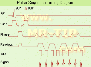
Echo Planar Imaging Timing Diagram (EPI) Echo planar imaging is one of the early magnetic resonance imaging sequences (also known as Intascan), used in applications like diffusion, perfusion, and functional magnetic resonance imaging. Other sequences acquire one k-space line at each phase encoding step. When the echo planar imaging acquisition strategy is used, the complete image is formed from a single data sample (all k-space lines are measured in one repetition time) of a gradient echo or spin echo sequence (see single shot technique) with an acquisition time of about 20 to 100 ms. The pulse sequence timing diagram illustrates an echo planar imaging sequence from spin echo type with eight echo train pulses. (See also Pulse Sequence Timing Diagram, for a description of the components.)
In case of a gradient echo based EPI sequence the initial part is very similar to a standard gradient echo sequence. By periodically fast reversing the readout or frequency encoding gradient, a train of echoes is generated.
EPI requires higher performance from the MRI scanner like much larger gradient amplitudes. The scan time is dependent on the spatial resolution required, the strength of the applied gradient fields and the time the machine needs to ramp the gradients.
In EPI, there is water fat shift in the phase encoding direction due to phase accumulations. To minimize water fat shift (WFS) in the phase direction fat suppression and a wide bandwidth (BW) are selected. On a typical EPI sequence, there is virtually no time at all for the flat top of the gradient waveform. The problem is solved by "ramp sampling" through most of the rise and fall time to improve image resolution.
The benefits of the fast imaging time are not without cost. EPI is relatively demanding on the scanner hardware, in particular on gradient strengths, gradient switching times, and receiver bandwidth. In addition, EPI is extremely sensitive to image artifacts and distortions.
spacer

• View the DATABASE results for 'Echo Planar Imaging' (19).Open this link in a new window


• View the NEWS results for 'Echo Planar Imaging' (1).Open this link in a new window.
 
Further Reading:
  Basics:
New Imaging Method Makes Brain Scans 7 Times Faster
Sunday, 9 January 2011   by www.dailytech.com    
Searchterm 'Diffusion Time' was also found in the following services: 
spacer
News  (2)  Resources  (1)  
 
Panorama 0.6TPanorama 0.2InfoSheet: - Devices -
Intro, 
Types of Magnets, 
Overview, 
etc.MRI Resource Directory:
 - Devices -
 
www.medical.philips.com/main/products/mri/products/panoramafamily/panorama0.23t_rt/features/ From Philips Medical Systems;
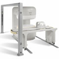
Panorama 0.6 T is the Philips Mid-Field Open MRI system. It is the most open MR scanner in the market, optimized for patient comfort and faster reconstruction time.
Device Information and Specification
CLINICAL APPLICATION
Whole body
CONFIGURATION
Open MRI/C-arm
Head, head-neck, extremity, neck, body/spine M-XL, shoulder, bilateral breast, wrist, TMJ, flex XS-S-M-L-XL-XXL
SYNCHRONIZATION
ECG/peripheral: Optional/yes, respiratory gating
PULSE SEQUENCES
SE, FE, IR, STIR, FFE, DEFFE, DESE, TSE, DETSE, Single shot SE, DRIVE, Balanced FFE, MRCP, Fluid Attenuated Inversion Recovery, Turbo FLAIR, IR-TSE, T1-STIR TSE, T2-STIR TSE, Diffusion Imaging, 3D SE, 3D FFE, Contrast Perfusion Analysis, MTC;; Angiography: CE-ANGIO, MRA 2D, 3D TOF
IMAGING MODES
Single, multislice, volume study, dynamic, SIMEX, multi chunk 3D, multiple stacks
TR
Min. 4.6 msec
TE
Min. 2.3 msec
SINGLE/MULTI SLICE
50 slices/sec
0.4 cm - 42 cm
1280 X 1024
MEASURING MATRIX
Up to 512 x 512
PIXEL INTENSITY
256 gray scale
MAGNET TYPE
Superconducting / iron core
Open x 47 cm x infinite (side-first patient entry)
MAGNET WEIGHT
38000 kg
H*W*D
254 x 244 x 325 cm
POWER REQUIREMENTS
400/480 V
COOLING SYSTEM TYPE
Liquid helium//air cool
0.00 L/hr helium
STRENGTH
20 mT/m
5-GAUSS FRINGE FIELD
2.4 m / 2.5 m
Passive/active
spacer

• View the DATABASE results for 'Panorama 0.6T™' (2).Open this link in a new window

MRI Resources 
DICOM - RIS - Jobs pool - Services and Supplies - Guidance - Portals
 
Panorama 0.23T™InfoSheet: - Devices -
Intro, 
Types of Magnets, 
Overview, 
etc.MRI Resource Directory:
 - Devices -
 
www.medical.philips.com/main/products/mri/products/panoramafamily/panorama0.23t_rt/features/ From Philips Medical Systems;
the Panorama 0.23 T, providing a new design optimized for patient comfort, faster reconstruction time than before (300 images/second) and new gradient specifications. Philips' Panorama 0.23 T I/T supports MR-guided interventions, resulting in minimally invasive procedures, more targeted surgery, reduced recovery time and shorter hospital stays. Optional OptoGuide functionality enables real-time needle tracking. Philips' Panorama 0.23 TPanorama 0.2 R/T is the first and only open MRI system to enable radiation therapy planning using MR data sets. The Panorama also features the new and consistent Philips User Interface, an essential element of the Vequion clinical IT family of products and services.
Device Information and Specification
CLINICAL APPLICATION
Whole body
CONFIGURATION
Open MRI/C-arm
Head, head-neck, extremity M-L, neck, body/spine S-XL, shoulder, bilateral breast, wrist, TMJ, flex XS-S-M-L-XL-XXL
SYNCHRONIZATION
ECG/peripheral: Optional/optional, respiratory gating
PULSE SEQUENCES
SE, FE, IR, FFE, DEFFE, DESE, TSE, DETSE, Single shot SE, DRIVE, Balanced FFE, MRCP, Fluid Attenuated Inversion Recovery, Turbo FLAIR, IR-TSE, T1-STIR TSE, T2-STIR TSE, Diffusion Imaging, 3D SE, 3D FFE, MTC;; Angiography: CE-ANGIO, MRA 2D, 3D TOF
IMAGING MODES
Single, multislice, volume study, dynamic, SIMEX, multi chunk 3D, multiple stacks
TR
Min. 6.2 msec
TE
Min. 2.8 msec
SINGLE/MULTI SLICE
50 slices/sec
0.4 cm - 40 cm
1280 X 1024
MEASURING MATRIX
Up to 512 x 512
PIXEL INTENSITY
256 gray scale
MAGNET TYPE
Resistive/iron core
Open x 46 cm x infinite (side-first patient entry)
MAGNET WEIGHT
13110 kg
H*W*D
196 x 121 x 176 cm
POWER REQUIREMENTS
400/480 V
COOLING SYSTEM TYPE
Closed loop chilled water (chiller included)
N/A
STRENGTH
19 mT/m
5-GAUSS FRINGE FIELD
2.4 m / 3.7 m
Passive/active
spacer

• View the DATABASE results for 'Panorama 0.23T™' (2).Open this link in a new window

 
Further Reading:
  News & More:
Magnetic resonance imaging guided musculoskeletal interventions at 0.23T: Chapter 4. Materials and methods
2002
MRI Resources 
Equipment - Collections - Coils - Cochlear Implant - Brain MRI - Anatomy
 
     1 - 5 (of 19)     next
Result Pages : [1]  [2 3 4]
 Random Page
 
Share This Page
FacebookTwitterLinkedIn

MR-TIP    
Community   
User
Pass
Forgot your UserID/Password ?    


Personalized protocols (age, gender, body habitus, etc.) lead to :
more automated planning 
improved patient comfort 
better diagnostics 
optimized image quality 
nothing 

Look
      Ups


MR-TIP.com uses cookies! By browsing MR-TIP.com, you agree to our use of cookies.

Magnetic Resonance - Technology Information Portal
Member of SoftWays' Medical Imaging Group - MR-TIP • Radiology-TIP • Medical-Ultrasound-Imaging • 
Copyright © 2003 - 2024 SoftWays. All rights reserved. [ 19 April 2024]
Terms of Use | Privacy Policy | Advertising
 [last update: 2024-02-26 03:41:00]