Magnetic Resonance - Technology Information Portal Welcome to MRI Technology
Info
  Sheets

Out-
      side


 'Circle of Willis' 
SEARCH FOR    
 
  2 3 5 A B C D E F G H I J K L M N O P Q R S T U V W X Y Z
Result : Searchterm 'Circle of Willis' found in 1 term [] and 4 definitions []
1 - 5 (of 5)     
Result Pages : [1]
Searchterm 'Circle of Willis' was also found in the following services: 
spacer
Resources  (3)  Forum  (2)  
 
Circle of WillisForum -
related threadsMRI Resource Directory:
 - Anatomy -
 
A large network of interconnecting blood vessels at the base of the brain that when visualized resembles a circle, the arteries effectively act as anastomoses for each other. This means that if any one of the communicating arteries becomes blocked, blood can flow from another part of the circle to ensure that blood flow is not compromised.
The circle of Willis is formed by both the internal carotid arteries, entering the brain from each side and the basilar artery, entering posteriorly. The connection of the vertebral arteries forms the basilar artery. The basilar artery divides into the right and left posterior cerebral arteries. The internal carotid arteries trifurcate into the anterior cerebral artery, middle cerebral artery, and posterior communicating artery. The two anterior cerebral arteries are joined together anteriorly by the anterior communicating artery. The posterior communicating arteries join the posterior cerebral arteries, completing the circle of Willis.
The time of flight angiography MRI technique allows imaging of the circle of Willis without the need of a contrast medium (best results with high field MRI). A cerebrovasular contrast enhanced magnetic resonance angiography (MRA) depicts the circle of Willis in addition to the vessels of the neck (carotid and vertebral arteries) with one bolus injection of a contrast agent.

For Ultrasound Imaging (USI) see Cerebrovascular Ultrasonography at Medical-Ultrasound-Imaging.com.
 
Images, Movies, Sliders:
 CE-MRA of the Carotid Arteries  Open this link in a new window
    
SlidersSliders Overview

 TOF-MRA Circle of Willis Inverted MIP  Open this link in a new window
    

 Circle of Willis, Time of Flight, MIP  Open this link in a new window
    
SlidersSliders Overview

 
spacer
 
• Share the entry 'Circle of Willis':  Facebook  Twitter  LinkedIn  
 
• Related Searches:
    • Angiography
    • Magnetic Resonance Angiography MRA
    • 3 Dimensional Magnetic Resonance Angiography
    • Time of Flight Angiography
    • Contrast Enhanced Magnetic Resonance Angiography
 
Further Reading:
  News & More:
Magnetic resonance angiography: current status and future directions
Wednesday, 9 March 2011   by www.jcmr-online.com    
Searchterm 'Circle of Willis' was also found in the following service: 
spacer
Ultrasound  (1) Open this link in a new window
3 Dimensional Magnetic Resonance AngiographyInfoSheet: - Sequences - 
Intro, 
Overview, 
Types of, 
etc.MRI Resource Directory:
 - MRA -
 
(3D MRA) The 3D angiography technique can be applied to focus on fast flowing (arterial) blood and to visualize small tortuous vessels. 3D TOF images are less sensitive to turbulent flow artifacts. The advantage of this approach is that the signal, acquired from the entire volume has an increased signal to noise ratio. Slices are defined by a second phase encoded axis, which divides the volume into 'partitions'. 3D TOF MRA is acquired with 3D FT slabs or multiple overlapping thin 3D FT slabs (MOTSA) depending on the coverage required and the range of flow-velocities under examination.
Such 3D techniques can provide equal spatial resolution along all three axes, i.e. be 'isotropic', or the partition thickness can be greater or less than the in plane spatial resolution in which case can be said to be 'anisotropic'. The circle of Willis, anatomy as well as its fast arterial flow, lends itself well to both 3D TOF and 2D or 3D phase contrast angiography.
 
Images, Movies, Sliders:
 CE MRA of the Aorta  Open this link in a new window
    
SlidersSliders Overview

 PCA-MRA 3D Brain Venography Colored MIP  Open this link in a new window
    

 CE-MRA of the Carotid Arteries Colored MIP  Open this link in a new window
    
SlidersSliders Overview

 Circle of Willis, Time of Flight, MIP  Open this link in a new window
    
SlidersSliders Overview

 
spacer

• View the DATABASE results for '3 Dimensional Magnetic Resonance Angiography' (2).Open this link in a new window

 
Further Reading:
  Basics:
CHAPTER 55: Ischemia
2003
Magnetic resonance angiography: current status and future directions
Wednesday, 9 March 2011   by www.jcmr-online.com    
MRI Resources 
Online Books - Jobs - MR Guided Interventions - MRCP - Contrast Enhanced MRI - MRI Training Courses
 
MAGNETOM Symphony™InfoSheet: - Devices -
Intro, 
Types of Magnets, 
Overview, 
etc.MRI Resource Directory:
 - Devices -
 
www.med.siemens.com/med/d/gg/mr/products/symphony1.html From Siemens Medical Systems;
with the introduction of this system, it is possible to perform contrast MR angiography for abdominal, thoracic and neck vessels from the origins to the circle of Willis. The system also has many newer features including functional imaging, spectroscopy, advanced body, ortho- and neuroimaging.
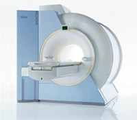
Device Information and Specification
CLINICAL APPLICATION
Whole body
CONFIGURATION
Compact
Yes/SVS or CSI opt.
SYNCHRONIZATION
ECG/peripheral, respiratory gating
PULSE SEQUENCES
GRE, IR, FIR, STIR, TrueIR/FISP, FSE, FLAIR, MT, SS-FSE, MT-SE, MTC, MSE, EPI, GMR, fat/water sat./exc.
IMAGING MODES
Single, multislice, volume study, multi angle, multi oblique
TR
2.4 msec std.; 2.0 opt.; 1.8 w/30 mT/m at 256matrix
TE
1.1 msec std.; 0.9 opt.; 0.78 w/30 mT/m at 256matrix
SINGLE/MULTI SLICE
178 images/sec at 256 x 256 at 100% FOV
0.5 cm - 50 cm
Min 2D/3D: 0.1/0.05 mm
1024 x 1024 full screen display
MEASURING MATRIX
64 x 64 to 1024 x 1024
PIXEL INTENSITY
21 micrometer in plane, 11 micrometer optional
60 cm
MAGNET WEIGHT
4050kg, 5500kg in operation
H*W*D
236 x 215 x 160 cm w/covers
POWER REQUIREMENTS
380/400/420/440/480 V
COOLING SYSTEM TYPE
Single cryogen, 2 stage refrig.
Liquid He
STRENGTH
20/35 mT/m standard, 30/52 opt.
5-GAUSS FRINGE FIELD
2.5 m / 4.0 m
Passive, act.; 1st order std./2nd opt.
spacer

• View the DATABASE results for 'MAGNETOM Symphony™' (2).Open this link in a new window

 
Further Reading:
  Basics:
Symphony Brochure(.pdf)
Searchterm 'Circle of Willis' was also found in the following services: 
spacer
Resources  (3)  Forum  (2)  
 
Multiple Overlapping Thin Slab (Slice) Acquisition
 
(MOTSA) This technique combines the best features of 2D time of flight angiography (2D TOF) and 3D TOF MRA. The MOTSA technique consists of multiple 2 cm thick 3D TOF slabs (which minimize saturation effects for through plane flow) combine to provide unlimited coverage similar to multiple 2D TOF slices. High resolution imaging of the carotid arteries is possible when image quality is of greater concern than acquisition time. Images with 1 mm (or less) spatial resolution in all three planes are required. The slabs typically overlap 25-40 to minimize the venetian blind artifact venetian blind artifact due to minimal saturation effects. MOTSA is an useful technique for the evaluation of vertebrobasilar ischemia and aneurysm scanning from the foramen magnum through the circle of Willis.
 
Images, Movies, Sliders:
 Circle of Willis, Time of Flight, MIP  Open this link in a new window
    
SlidersSliders Overview

 
spacer
Searchterm 'Circle of Willis' was also found in the following service: 
spacer
Ultrasound  (1) Open this link in a new window
Time of Flight AngiographyInfoSheet: - Sequences - 
Intro, 
Overview, 
Types of, 
etc.MRI Resource Directory:
 - MRA -
 
(TOF) The time of flight angiography is used for the imaging of vessels. Usually the sequence type is a gradient echo sequences with short TR, acquired with slices perpendicular to the direction of blood flow.
The source of diverse flow effects is the difference between the unsaturated and presaturated spins and creates a bright vascular image without the invasive use of contrast media. Flowing blood moves unsaturated spins from outside the slice into the imaging plane. These completely relaxed spins have full equilibrium magnetization and produce (when entering the imaging plane) a much higher signal than stationary spins if a gradient echo sequence is generated. This flow related enhancement is also referred to as entry slice phenomenon, or inflow enhancement.
Performing a presaturation slab on one side parallel to the slice can selectively destroy the MR signal from the in-flowing blood from this side of the slice. This allows the technique to be flow direction sensitive and to separate arteriograms or venograms. When the local magnetization of moving blood is selectively altered in a region, e.g. by selective excitation, it carries the altered magnetization with it when it moves, thus tagging the selected region for times on the order of the relaxation times.
For maximum flow signal, a complete new part of blood has to enter the slice every repetition (TR) period, which makes time of flight angiography sensitive to flow-velocity. The choice of TR and slice thickness should be appropriate to the expected flow-velocities because even small changes in slice thickness influences the performance of the TOF sequence. The use of sequential 2 dimensional Fourier transformation (2DFT) slices, 3DFT slabs, or multiple 3D slabs (chunks) are depending on the coverage required and the range of flow-velocities.
3D TOF MRA is routinely used for evaluating the Circle of Willis.

See also Magnetic Resonance Angiography and Contrast Enhanced Magnetic Resonance Angiography.
 
Images, Movies, Sliders:
 TOF-MRA Circle of Willis Inverted MIP  Open this link in a new window
    

 Circle of Willis, Time of Flight, MIP  Open this link in a new window
    
SlidersSliders Overview

 
Radiology-tip.comradCT Angiography,  Coronary Angiogram
spacer
Medical-Ultrasound-Imaging.comColor Power Angio,  Doppler Ultrasound
spacer

• View the DATABASE results for 'Time of Flight Angiography' (11).Open this link in a new window

 
Further Reading:
  Basics:
MR–ANGIOGRAPHY(.pdf)
  News & More:
Magnetic resonance angiography: current status and future directions
Wednesday, 9 March 2011   by www.jcmr-online.com    
MRI Resources 
Nerve Stimulator - MR Guided Interventions - Pathology - Movies - Safety Training - Safety Products
 
     1 - 5 (of 5)     
Result Pages : [1]
 Random Page
 
Share This Page
FacebookTwitterLinkedIn

MR-TIP    
Community   
User
Pass
Forgot your UserID/Password ?    


New acceleration techniques will :
reduce scan times 
cause artifacts 
increase expenses 
be useful if you have a lot of experience 
doesn't do much 
never heard of 

Look
      Ups


MR-TIP.com uses cookies! By browsing MR-TIP.com, you agree to our use of cookies.

Magnetic Resonance - Technology Information Portal
Member of SoftWays' Medical Imaging Group - MR-TIP • Radiology-TIP • Medical-Ultrasound-Imaging • 
Copyright © 2003 - 2024 SoftWays. All rights reserved. [ 19 April 2024]
Terms of Use | Privacy Policy | Advertising
 [last update: 2024-02-26 03:41:00]