| Info
Sheets |
| | | | | | | | | | | | | | | | | | | | | | | | |
 | Out-
side |
| | | | |
|
| | | | |
Result : Searchterm 'Brain MRI' found in 1 term [ ] and 13 definitions [ ] and 13 definitions [ ], (+ 14 Boolean[ ], (+ 14 Boolean[ ] results ] results
| | previous 16 - 20 (of 28) nextResult Pages : [1] [2 3] [4 5 6] |  | |  | Searchterm 'Brain MRI' was also found in the following services: | | | | |
| |  |
| |
|

From GE Healthcare;
'EXCITE technology has the potential to open the door to new imaging techniques and clinical applications, leaping beyond conventional two and three-dimensional MRI to true 4D imaging that will improve the diagnosis of disease throughout the human body from head to foot.' Robert R. Edelman, M.D., Professor of Radiology at Northwestern University Medical School and Chairman, Department of Radiology, at Evanston Northwestern Healthcare.
Device Information and Specification CLINICAL APPLICATION Whole body Head and body coil standard; all other coils optional; open architecture makes system compatible with a wide selection of coils Optional 2D/3D brain and prostate Standard: SE, IR, 2D/3D GRE and SPGR, Angiography: 2D/3D TOF, 2D/3D Phase Contrast;; 2D/3D FSE, 2D/3D FGRE and FSPGR, SSFP, FLAIR, EPI, optional: 2D/3D Fiesta, FGRET, Spiral, TensorTR 1.3 to 12000 msec in increments of 1 msec TE 0.4 to 2000 msec in increments of 1 msec 2D 0.7 mm to 20 mm; 3D 0.1 mm to 5 mm 128x512 steps 32 phase encode 0.08 mm; 0.02 mm optional POWER REQUIREMENTS 480 or 380/415 less than 0.03 L/hr liquid heliumSTRENGTH SmartSpeed 23 mT/m, HiSpeed Plus 33 mT/m, EchoSpeed Plus 33 mT/m 4.0 m x 2.8 m axial x radial | | | | | |
| | | | | |
| |
|

From GE Healthcare;
The Signa HDx MRI system is GE's leading edge whole body magnetic resonance scanner designed to support high resolution, high signal to noise ratio, and short scan times.
Signa HDx 3.0T offers new technologies like ultra-fast image reconstruction through the new XVRE recon engine, advancements in parallel imaging algorithms and the broadest range of premium applications. The HD applications, PROPELLER (high-quality brain imaging extremely resistant to motion artifacts), TRICKS (contrast-enhanced angiographic vascular lower leg imaging), VIBRANT (for breast MRI), LAVA (high resolution liver imaging with shorter breath holds and better organ coverage) and MR Echo (high-definition cardiac images in real time) offer unique capabilities.
Device Information and Specification CLINICAL APPLICATION Whole body
CONFIGURATION Compact short bore SE, IR, 2D/3D GRE, RF-spoiled GRE, 2DFGRE, 2DFSPGR, 3DFGRE, 3DFSPGR, 3DTOFGRE, 3DFSPGR, 2DFSE, 2DFSE-XL, 2DFSE-IR, T1-FLAIR, SSFSE, EPI, DW-EPI, BRAVO, Angiography: 2D/3D TOF, 2D/3D phase contrast vascular IMAGING MODES Single, multislice, volume study, fast scan, multi slab, cine, localizer H*W*D 240 x 2216,6 x 201,6 cm POWER REQUIREMENTS 480 or 380/415, 3 phase ||
COOLING SYSTEM TYPE Closed-loop water-cooled grad. | | | | | |
| | | | | |
| |
|

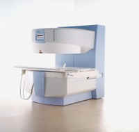
From GE Healthcare;
three dedicated MRI systems - a neuro imager, a cardiovascular system, and a whole body scanner - all in one system.
Device Information and Specification CLINICAL APPLICATION Whole body Head and body coil standard; all other coils optional; open architecture makes system compatible with a wide selection of coils Optional 2D/3D brain and prostate Standard: SE, IR, 2D/3D GRE and SPGR, Angiography: 2D/3D TOF, 2D/3D Phase Contrast;; 2D/3D FSE, 2D/3D FGRE and FSPGR, SSFP, FLAIR, EPI, optional: 2D/3D Fiesta, FGRET, Spiral, TensorTR 1.2 to 12000 msec in increments of 1 msec TE 0.4 to 2000 msec in increments of 1 msec 2D 0.7 mm to 20 mm; 3D 0.1 mm to 5 mm 128x512 steps 32 phase encode POWER REQUIREMENTS 480 or 380/415 Less than 0.03 L/hr liquid He STRENGTH Zoom 40 mT/m, whole body 23 mT/m 4.0 m x 2.8 m axial x radial | | | |
• View the DATABASE results for 'Signa Infinity 1.5T™ TwinSpeed with Excite' (2).
| | | | |  Further Reading: Further Reading: | News & More:
|
|
| |
| | | Searchterm 'Brain MRI' was also found in the following services: | | | | |
| | |
| |
|
Device Information and Specification
CLINICAL APPLICATION
Whole body
GRE, IR, FIR, STIR, TrueIR/FISP, FSE, MT, SS-FSE, MT-SE, MTC, MSE, GMR, fat/water sat./exc.
IMAGING MODES
Single, multislice, volume study, multi angle
512 x 512 full screen display
POWER REQUIREMENTS
380/400/420/440/480 V
| | | |
• View the DATABASE results for 'MAGNETOM Concerto™' (2).
| | | | |
| | | | | |
| |
|
Flow phenomena are intrinsic processes in the human body. Organs like the heart, the brain or the kidneys need large amounts of blood and the blood flow varies depending on their degree of activity. Magnetic resonance imaging has a high sensitivity to flow and offers accurate, reproducible, and noninvasive methods for the quantification of flow. MRI flow measurements yield information of blood supply of of various vessels and tissues as well as cerebro spinal fluid movement.
Flow can be measured and visualized with different pulse sequences (e.g. phase contrast sequence, cine sequence, time of flight angiography) or contrast enhanced MRI methods (e.g. perfusion imaging, arterial spin labeling).
The blood volume per time (flow) is measured in: cm3/s or ml/min. The blood flow-velocity decreases gradually dependent on the vessel diameter, from approximately 50 cm per second in arteries with a diameter of around 6 mm like the carotids, to 0.3 cm per second in the small arterioles.
Different flow types in human body:
•
Behaves like stationary tissue, the signal intensity depends on T1, T2 and PD = Stagnant flow
•
Flow with consistent velocities across a vessel = Laminar flow
•
Laminar flow passes through a stricture or stenosis (in the center fast flow, near the walls the flow spirals) = Vortex flow
•
Flow at different velocities that fluctuates = Turbulent flow
See also Flow Effects, Flow Artifact, Flow Quantification, Flow Related Enhancement, Flow Encoding, Flow Void, Cerebro Spinal Fluid Pulsation Artifact, Cardiovascular Imaging and Cardiac MRI. | | | | | |
• View the DATABASE results for 'Flow' (113).
| | |
• View the NEWS results for 'Flow' (7).
| | | | | | Further Reading: | News & More:
|
|
| |
| | | | |
| | |
| | | |
|
| |
 | Look
Ups |
| |