 | Info
Sheets |
| | | | | | | | | | | | | | | | | | | | | | | | |
 | Out-
side |
| | | | |
|
| | | | |
Result : Searchterm 'Axial' found in 2 terms [ ] and 18 definitions [ ] and 18 definitions [ ] ]
| | previous 6 - 10 (of 20) nextResult Pages : [1] [2 3 4] |  | |  | Searchterm 'Axial' was also found in the following services: | | | | |
| |  |
| |
|
Cervical spine MRI is a suitable tool in the assessment of all cervical spine (vertebrae C1 - C7) segments (computed tomography (CT) images may be unsatisfactory close to the thoracic spine due to shoulder artifacts). The cervical spine is particularly susceptible to degenerative problems caused by the complex anatomy and its large range of motion.
Advantages of magnetic resonance imaging MRI are the high soft tissue contrast (particularly important in diagnostics of the spinal cord), the ability to display the entire spine in sagittal views and the capacity of 3D visualization. Magnetic resonance myelography is a useful supplement to conventional MRI examinations in the investigation of cervical stenosis. Myelographic sequences result in MR images with high contrast that are similar in appearance to conventional myelograms. Additionally, open MRI studies provide the possibility of weight-bearing MRI scan to evaluate structural positional and kinetic changes of the cervical spine. Indications of cervical spine MRI scans include the assessment of soft disc herniations, suspicion of disc hernia recurrence after operation, cervical spondylosis, osteophytes, joint arthrosis, spinal canal lesions (tumors, multiple sclerosis, etc.), bone diseases (infection, inflammation, tumoral infiltration) and paravertebral spaces.
State-of-the-art phased array spine coils and high performance MRI machines provide high image quality and short scan time. Imaging protocols for the cervical spine includes sagittal T1 weighted and T2 weighted sequences with 3-4 mm slice thickness and axial slices; usually contiguous from C2 through T1. Additionally, T2 fat suppressed and T1 post contrast images are often useful in spine imaging. See also Lumbar Spine MRI.
| | | | | | | | | | |  Further Reading: Further Reading: | News & More:
|
|
| |
| | | Searchterm 'Axial' was also found in the following services: | | | | |
| | |
| |
|
This term is commonly used for a particular kind of gradient coil, commonly used to create magnetic field gradients perpendicular to the main magnetic field. A golay coil (a special kind of saddle coils) produces a linear gradient in the x and y axes that requires wires running along the bore of the magnet. Such a coil produces a very linear field, but the linearity is lost rapidly away from the central plane. A number of pairs with different axial separations can be used to improve this. | | | |
• View the DATABASE results for 'Golay Coil' (3).
| | | | |
| | | | | |
| |
|
Knee MRI, with its high soft tissue contrast is one of the main imaging tools to depict knee joint pathology. MRI allows accurate imaging of intra-articular structures such as ligaments, cartilage, menisci, bone marrow, synovium, and adjacent soft tissue.
Knee exams require a dedicated extremity coil, providing a homogenous imaging volume and high SNR to ensure best signal coverage.
A complete knee MR examination includes for example sagittal and coronal T1 weighted, and proton density weighted pulse sequences +/- fat saturation, or STIR sequences. For high spatial resolution, maximal 4 mm thick slices with at least an in plane resolution of 0.75 mm and small gap are recommended. To depict the anterior cruciate ligament clearly, the sagittal plane has to be rotated 10 - 20° externally (parallel to the medial border of the femoral condyle). Retropatellar cartilage can bee seen for example in axial T2 weighted gradient echo sequences with Fatsat. However, the choice of the pulse sequences is depended of the diagnostic question, the used scanner, and preference of the operator.
Diagnostic quality in knee imaging is possible with field strengths ranging from 0.2 to 3T. With low field strengths more signal averages must be measured, resulting in increased scan times to provide equivalent quality as high field strengths.
More diagnostic information of meniscal tears and chondral defects can be obtained by direct magnetic resonance arthrography, which is done by introducing a dilute solution of gadolinium in saline (1:1000) into the joint capsule. The knee is then scanned in all three planes using T1W sequences with fat suppression. For indirect arthrography, the contrast is given i.v. and similar scans are started 20 min. after injection and exercise of the knee.
Frequent indications of MRI scans in musculoskeletal knee diseases are: e.g., meniscal degeneration and tears, ligament injuries, osteochondral fractures, osteochondritis dissecans, avascular bone necrosis and rheumatoid arthritis. See also Imaging of the Extremities and STIR. | | | | | |
• View the DATABASE results for 'Knee MRI' (4).
| | |
• View the NEWS results for 'Knee MRI' (4).
| | | | | | Further Reading: | | Basics:
|
|
News & More:
| |
| |
| | | Searchterm 'Axial' was also found in the following services: | | | | |
| | |
| |
|
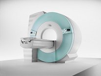
From Siemens Medical Systems;
70 cm + 125 cm + 1.5T and Tim - a combination never seen before in MRI ...
MAGNETOM Espree™s unique open bore design can accommodate more types of patients than other 1.5T systems on the market today, in particular the growing population of obese patients. The power of 1.5T combined with Tim technology boosts signal to noise, which is necessary to adequately image obese patients.
Device Information and Specification
CLINICAL APPLICATION
Whole body
Body, Tim [32 x 8], Tim [76 coil elements with up to 18 RF channels])
GRE, IR, FIR, STIR, TrueIR/FISP, FSE, FLAIR, MT, SS-FSE, MT-SE, MTC, MSE, EPI, 3D DESS//CISS/PSIF, GMR
IMAGING MODES
Single, multislice, volume study, multi angle, multi oblique
Image Processor reconstructing up to 3226 images per second (256 x 256, 25% recFoV)
1024 x 1024 full screen display
| | | |
• View the DATABASE results for 'MAGNETOM Espree™' (2).
| | | | | | Further Reading: | News & More:
|
|
| |
| | | Searchterm 'Axial' was also found in the following services: | | | | |
| | |
| |
|

From ONI Medical Systems, Inc.;
MSK-Extreme™ MRI system is a dedicated high field extremity imaging device, designed to provide orthopedic surgeons and other physicians with detailed diagnostic images of the foot, ankle, knee, hand, wrist and elbow, all with the clinical confidence and advantages derived from high field, whole body MRI units. The light weight (less than 650 kg) of the OrthOne System performs rapid patient studies, is easy to operate, has a patient friendly open environment and can be installed in a practice office or hospital, all at a cost similar to a low field extremity machine.
New features include a more powerful operating system that offers increased scan speed as well as a 160-mm knee coil with higher signal to noise ratio, and the option of a CD burner.
Device Information and Specification 16 cm knee, 18 cm lower extremity;; 12.3 cm upper extremity, additional high resolution v-SPEC Coils: 80 mm, 100 mm, or 145 mm. SE, FSE, GE2D, GE3D, Inversion recovery (IR), Driven Equilibrium, Fat Saturation (FS), STIR, MT, PD, Flow Compensation (FC), RF spoiling, MTE, No Phase Wrap (NPW) IMAGING MODES Scout, single, multislice, volume 2D less than 200 msec/image X/Y: 64-512; 2 pixel steps 4,096 grey lvls; 256 lvls in 3D POWER REQUIREMENTS 115VAC, 1phase, 20A; 208VAC, 3 phase, 30A COOLING SYSTEM TYPE LHe with 2 stage cold head 1.25m radial x 1.8m axial | | | | | | | Further Reading: | Basics:
|
|
| |
| | | | |
| | | |
|
| |
 | Look
Ups |
| |


