 | Info
Sheets |
| | | | | | | | | | | | | | | | | | | | | | | | |
 | Out-
side |
| | | | |
|
| | | | | |  | Searchterm 'Angiography' was also found in the following services: | | | | |
|  |  |
| |
|

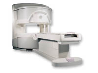
From GE Healthcare;
three dedicated MRI systems - a neuro imager, a cardiovascular system, and a whole body scanner - all in one system.
Device Information and Specification CLINICAL APPLICATION Whole body Head and body coil standard; all other coils optional; open architecture makes system compatible with a wide selection of coils Optional 2D/3D brain and prostate Standard: SE, IR, 2D/3D GRE and SPGR, Angiography: 2D/3D TOF, 2D/3D Phase Contrast;; 2D/3D FSE, 2D/3D FGRE and FSPGR, SSFP, FLAIR, EPI, optional: 2D/3D Fiesta, FGRET, Spiral, TensorTR 1.2 to 12000 msec in increments of 1 msec TE 0.4 to 2000 msec in increments of 1 msec 2D 0.7 mm to 20 mm; 3D 0.1 mm to 5 mm 128x512 steps 32 phase encode POWER REQUIREMENTS 480 or 380/415 Less than 0.03 L/hr liquid He STRENGTH Zoom 40 mT/m, whole body 23 mT/m 4.0 m x 2.8 m axial x radial | | | | | |  Further Reading: Further Reading: | News & More:
|
|
| |
| | | Searchterm 'Angiography' was also found in the following services: | | | | |
| | |
| |
|

From GE Healthcare;
'EXCITE technology has the potential to open the door to new imaging techniques and clinical applications, leaping beyond conventional two and three-dimensional MRI to true 4D imaging that will improve the diagnosis of disease throughout the human body from head to foot.' Robert R. Edelman, M.D., Professor of Radiology at Northwestern University Medical School and Chairman, Department of Radiology, at Evanston Northwestern Healthcare.
Device Information and Specification CLINICAL APPLICATION Whole body Head and body coil standard; all other coils optional; open architecture makes system compatible with a wide selection of coils Optional 2D/3D brain and prostate Standard: SE, IR, 2D/3D GRE and SPGR, Angiography: 2D/3D TOF, 2D/3D Phase Contrast;; 2D/3D FSE, 2D/3D FGRE and FSPGR, SSFP, FLAIR, EPI, optional: 2D/3D Fiesta, FGRET, Spiral, TensorTR 1.3 to 12000 msec in increments of 1 msec TE 0.4 to 2000 msec in increments of 1 msec 2D 0.7 mm to 20 mm; 3D 0.1 mm to 5 mm 128x512 steps 32 phase encode 0.08 mm; 0.02 mm optional POWER REQUIREMENTS 480 or 380/415 less than 0.03 L/hr liquid heliumSTRENGTH SmartSpeed 23 mT/m, HiSpeed Plus 33 mT/m, EchoSpeed Plus 33 mT/m 4.0 m x 2.8 m axial x radial | | | |
• View the DATABASE results for 'Signa Infinity 1.5T™ with Excite' (2).
| | | | |
| | | | | |
| |
|
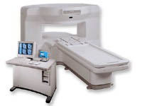
From GE Healthcare;
a friendly and less confining appearance targets the 7% of individuals who refuse to have an MRI because of claustrophobia. This open MRI system is also up to three times faster than other scanners, therefore the Signa OpenSpeed™ reducing exam time and scheduling
issues. In addition, a swing table provides better access and supports up to 500 pounds.
Device Information and Specification CLINICAL APPLICATION Whole body Standard: SE, IR, 2D/3D GRE and SPGR, Angiography: 2D/3D TOF, 2D/3D Phase Contrast;; 2D/3D FSE, 2D/3D FGRE and FSPGR, SSFP, FLAIR, EPI, optional: 2D/3D Fiesta, FGRET, Spiral, TensorTR 1.3 to 12000 msec in increments of 1 msec TE 0.4 to 2000 msec in increments of 1 msec 2D: 0.8mm - 20mm 3D: 0.1mm - 20mm 0.08 mm; 0.02 mm optional POWER REQUIREMENTS 200 - 480, 3-phase | | | |
• View the DATABASE results for 'Signa OpenSpeed™' (2).
| | | | | | Further Reading: | News & More:
|
|
| |
| | | Searchterm 'Angiography' was also found in the following services: | | | | |
| | |
| |
|
From GE Healthcare;
the New Signa Profile/i is a patient friendly open MRI system that virtually eliminates patient anxiety and claustrophobia, without compromising diagnostic utility.
Device Information and Specification CLINICAL APPLICATION Whole body Integrated transmit body coil, body flex sizes: M, L, XL, quadrature, head coil quadrature, 4 channel neurovascular array, 8 channel CTL array, quad. c- spine, 2 channel shoulder array, extremity coil, 3 channel wrist array, 4 channel breast array, 6, 9, 11 inch general purpose loop coils Standard: SE, IR, 2D/3D GRE and SPGR, Angiography: 2D/3D TOF, 2D/3D phase contrast; 2D/3D FSE, 2D/3D FRFSE, FGRE and FSPGR, SSFP, FLAIR, EPI, optional: 2D/3D Fiesta, fat/water separation, T1 FLAIRIMAGING MODES Localizer, single slice, multislice, volume, fast, POMP, multi slab, cine, slice and frequency zip, extended dynamic range, tailored RF TR 6 to 12000 msec in increments of 1 msec TE 1.3 to 2000 msec in increments of 1 msec 2D: 2.7mm - 20mm 3D: 0.2mm - 5mm 0.08 mm; 0.02 mm optional 10,000 kg w/gradient enclosure POWER REQUIREMENTS 200 - 480, 3-phase COOLING SYSTEM TYPE None required | | | |
• View the DATABASE results for 'Signa Profile™' (2).
| | | | |
| | | Searchterm 'Angiography' was also found in the following services: | | | | |
| | |
| |
|
Sinerem® is the brand name (same as Combidex®) for an ultrasmall superparamagnetic iron oxide ( USPIO) to detect metastatic disease in lymph nodes. Metastatic nodes show less uptake of this MRI contrast agent, which results in less signal decrease and allows the differentiation of normal lymph nodes from normal-sized, metastatic nodes.
Lymph node imaging with Sinerem® is performed 24 to 36 hours after slow infusion. Normal lymph nodes turn black post contrast, namely on T2* weighted images. Metastatic lymph nodes remain unchanged in signal intensity.
Indication and Diseases: Cancer, Imaging for diagnosis, Lymphatic disorders.
See Ferumoxtran, and Classifications, Characteristics, etc.
Guerbet decided in 2007 to withdraw its Marketing Authorisation Application
(MAA) for Sinerem.Drug Information and Specification r1=25, r2=160, B0=0.47T, r1=23.3, r2=48.9, B0=0.47T PHARMACOKINETIC Vascular, lymph v. hepatocyte (AG-USPIO) PREPARATION Suspend in an isotonic glucose solution DO NOT RELY ON THE INFORMATION PROVIDED HERE, THEY ARE
NOT A SUBSTITUTE FOR THE ACCOMPANYING PACKAGE INSERT! | | | |
• View the DATABASE results for 'Sinerem®' (6).
| | | | | | Further Reading: | | Basics:
|
|
News & More:
| |
| |
| | | | |
| | |
| | | |
|
| |
 | Look
Ups |
| |


