| Info
Sheets |
| | | | | | | | | | | | | | | | | | | | | | | | |
 | Out-
side |
| | | | |
|
| | | | |
Result : Searchterm '3t' found in 1 term [ ] and 16 definitions [ ] and 16 definitions [ ] ]
| | 1 - 5 (of 17) nextResult Pages : [1] [2 3 4] |  | |  | Searchterm '3t' was also found in the following services: | | | | |
| |  |
| |
|

From Philips Medical Systems;
the Panorama 0.23 T, providing a new design optimized for patient comfort, faster reconstruction time than before (300 images/second) and new gradient
specifications. Philips' Panorama 0.23 T I/T supports MR-guided interventions, resulting in minimally invasive procedures, more targeted surgery, reduced recovery time and shorter hospital stays. Optional OptoGuide functionality enables real-time needle tracking. Philips' Panorama 0.23 TPanorama 0.2 R/T is the first and only open MRI system to enable radiation therapy planning using MR data sets. The Panorama also features the new and consistent Philips User Interface, an essential element of the Vequion clinical IT family of products and services.
Device Information and Specification CLINICAL APPLICATION Whole body SE, FE, IR, FFE, DEFFE, DESE, TSE, DETSE, Single shot SE, DRIVE, Balanced FFE, MRCP, Fluid Attenuated Inversion Recovery, Turbo FLAIR, IR-TSE, T1-STIR TSE, T2-STIR TSE, Diffusion Imaging, 3D SE, 3D FFE, MTC;; Angiography: CE-ANGIO, MRA 2D, 3D TOFOpen x 46 cm x infinite (side-first patient entry) POWER REQUIREMENTS 400/480 V COOLING SYSTEM TYPE Closed loop chilled water ( chiller included) | | | | | | • Share the entry 'Panorama 0.23T™':    | | | | |  Further Reading: Further Reading: | News & More:
|
|
| |
| | | | |  |
| |
|
The principal advantage of MRI at high field is the increase in signal to noise ratio. This can be used to improve anatomic and/or temporal resolution and reduce scan time while preserving image quality. MRI devices for whole body imaging for human use are available up to 3 tesla (3T). Functional MRI ( fMRI) and MR spectroscopy ( MRS) benefit significantly. In addition, 3T machines have a great utility in applications such as TOF MRA and DTI. Higher field strengths are used for imaging of small parts of the body or scientific animal experiments. Higher contrast may permit reduction of gadolinium doses and, in some cases, earlier detection of disease.
Using high field MRI//MRS, the RF-wavelength and the dimension of the human body complicating the development of MR coils. The absorption of RF power causes heating of the tissue. The energy deposited in the patient's tissues is fourfold higher at 3T than at 1.5T. The specific absorption rate (SAR) induced temperature changes of the human body are the most important safety issue of high field MRI//MRS.
Susceptibility and chemical shift dispersion increase like T1, therefore high field MRI occasionally exhibits imaging artifacts. Most are obvious and easily recognized but some are subtle and mimic diseases. A thorough understanding of these artifacts is important to avoid potential pitfalls. Some imaging techniques or procedures can be utilized to remove or identify artifacts. See also Diffusion Tensor Imaging.
See also the related poll result: ' In 2010 your scanner will probably work with a field strength of' | | | | | |
• View the DATABASE results for 'High Field MRI' (16).
| | |
• View the NEWS results for 'High Field MRI' (9).
| | | | | | Further Reading: | | Basics:
|
|
News & More:
| |
| |
| | | | | |
| |
|
Device Information and Specification CLINICAL APPLICATION Whole body SE, IR, 2D/3D TurboSE, Turbo IR, Dark-Fluid IR, True IR, 2D/3D MEDIC, 2D/3D GRE FLASH, 2D/3D GRE FISP, 2D/3D PSIF, 2D TurboFLASH, 3D MP-RAGE, 3D TurboFLASH, 2D/3D TOF angiography, MTC, TONE with 3D TOF MRA, GMR, LOTA IMAGING MODES Single, multislice, volume study, multi angle, multi oblique178 images/sec at 256 x 256 at 100% FOV1024 x 1024 full screen display POWER REQUIREMENTS 380/400/420/440/480 V Passive, act.; 1st order std./2nd opt. | | | | | |
| | | Searchterm '3t' was also found in the following services: | | | | |
| | |
| |
|

Philips Medical System is the diagnostics business of Royal Philips Electronics of the Netherlands, one of the world's biggest electronics companies and Europe's largest. Philips is quoted on the NYSE (symbol: PHG), London, Frankfurt, Amsterdam and other stock exchanges.
On October 19, 2001, Philips Medical Systems completed a 3-year acquisition strategy through its purchase of Marconi Medical Systems. Marconi Medical Systems offered leading multislice CT, MRI, and Nuclear Gamma Camera systems to medical institutions around the world. As well as new 3.0T developments, Philips is also in collaboration with researchers at the University of Nottingham, with the intention of developing an ultrahigh field strength clinical 7.0T whole body MR system.
MRI Scanners:
0.2 3T to 1.0T:
1.5T:
3.0T to 7.0T:
Hybrid Scanners:
Contact Information MAIL Philips Medical Systems
3000 Minuteman Road
Andover, MA 01810-1099
USA | | | |
• View the DATABASE results for 'Philips Medical Systems' (14).
| | |
• View the NEWS results for 'Philips Medical Systems' (12).
| | | | | | Further Reading: | News & More:
|
|
| |
| | | | | |
| |
|

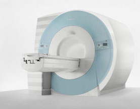
The range of diagnostics and imaging systems of Siemens Medical Systems covers ultrasound, nuclear medicine, angiography, magnetic resonance, computer tomography and patient monitoring. Siemens is one of the three leading MRI manufacturers, which together account for approximately 80 percent of the MRI machines installed worldwide. Siemens currently offers the Allegra 3T MRI, which is for head scanning only, but the company will also be launching the Trio MRI, a 3T whole body scanner.
Siemens has formed partnerships with more than ten research institutions and private practitioners to define a comprehensive MRI examination and compare MR to currently established cardiovascular modalities, thereby defining optimal diagnosis and treatment.
MRI Scanners:
0.2T to 1.0T:
1.5T:
3.0T to 7.0T:
Hybrid Scanners:
Mobile Solutions:
•
MAGNETOM Espree 1.5T, MAGNETOM Avanto 1.5T and MAGNETOM ESSENZA 1.5T are also offered by Siemens on certified trailers.
Contact Information MAIL
Siemens Medical Solutions
Health Services Corporation
51 Valley Stream Parkway
Malvern, PA 19355
USA | | | |
• View the DATABASE results for 'Siemens Medical Systems' (14).
| | |
• View the NEWS results for 'Siemens Medical Systems' (3).
| | | | | | Further Reading: | | Basics:
|
|
News & More:
| |
| |
| | | | |
| | | 1 - 5 (of 17) nextResult Pages : [1] [2 3 4] |
| |
|
| |
 | Look
Ups |
| |