Magnetic Resonance - Technology Information Portal Welcome to MRI Technology
Info
  Sheets

Out-
      side


 MRI Database 
SEARCH FOR    
  2 3 5 A B C D E F G H I J K L M N O P Q R S T U V W X Y Z
previous     61 - 65 (of 98)     next 
Fa-Fa   Fa-Fa   Fa-Fa   Fa-Fa   Fa-Fe   Fe-Fe   Fe-Fe   Fe-Fe   Fe-Fi   Fi-Fi   Fi-Fi   Fi-Fl   FL-Fl     Fl-Fl   Fl-Fl   Fo-Fo   FO-Fr   Fr-Fr   Fr-Fu   Fu-Fu   
MRI Resources 
Movies - Case Studies - Societies - Brain MRI - PACS - Stent
 
FLEXART™InfoSheet: - Devices -
Intro, 
Types of Magnets, 
Overview, etc.MRI Resource Directory:
 - Devices -
 
From Toshiba America Medical Systems Inc.; www3.toshiba.co.jp/medical/products/mr/flex/index.htm 
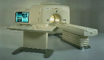
FLEXART™ series is a 0.5 T superconducting MRI system that has been designed to meet the expanding role of MRI in today's clinical environment. The system utilizes innovative technologies such as digital RF, high speed actively shielded gradients and optimized RF coils which support a wide range of MRI developments.
Device Information and Specification
CLINICAL APPLICATION
Whole body
CONFIGURATION
Compact
Quadrature, solenoid and multi-channel configurations
SYNCHRONIZATION
ECG/peripheral optional, respiratory gating
PULSE SEQUENCES
SE, FE, IR, FastSE, FastIR, FastFLAIR, Fast STIR, FastFE, FASE, Hybrid EPI, Multi Shot EPI; Angiography: 2D(gate/non-gate)/3D TOF, SORS-STC
IMAGING MODES
Single, multislice, volume study
POWER REQUIREMENTS
380/400/415/440/480 V
COOLING SYSTEM TYPE
Closed-loop water-cooled
STRENGTH
25 mT/m
Passive, active
spacer

• View the DATABASE results for 'FLEXART™' (2).Open this link in a new window

Flip Angle
 
(FA) The flip angle a is used to define the angle of excitation for a field echo pulse sequence. It is the angle to which the net magnetization is rotated or tipped relative to the main magnetic field direction via the application of a RF excitation pulse at the Larmor frequency. It is also referred to as the tip angle, nutation angle or angle of nutation.
The radio frequency power (which is proportional to the square of the amplitude) of the pulse is proportional to a through which the spins are tilted under its influence. Flip angles between 0° and 90° are typically used in gradient echo sequences, 90° and a series of 180° pulses in spin echo sequences and an initial 180° pulse followed by a 90° and a 180° pulse in inversion recovery sequences.
spacer

• View the NEWS results for 'Flip Angle' (1).Open this link in a new window.

• View the DATABASE results for 'Flip Angle' (37).Open this link in a new window

 
Further Reading:
  Basics:
What MRI Sequences Produce the Highest Specific Absorption Rate (SAR), and Is There Something We Should Be Doing to Reduce the SAR During Standard Examinations?
Thursday, 16 April 2015   by www.ajronline.org    
Mapping of low flip angles in magnetic resonance(.pdf)
Saturday, 1 January 2011   by www.hal.inserm.fr    
  News & More:
A practical guideline for T1 reconstruction from various flip angles in MRI
Saturday, 1 October 2016   by journals.sagepub.com    
Clinical evaluation of a speed optimized T2 weighted fast spin echo sequence at 3.0 T using variable flip angle refocusing, half-Fourier acquisition and parallel imaging
Wednesday, 25 October 2006
MRI Resources 
Sequences - Spectroscopy pool - Functional MRI - MR Guided Interventions - Patient Information - Anatomy
 
FlowForum -
there are related threads
 
Flow phenomena are intrinsic processes in the human body. Organs like the heart, the brain or the kidneys need large amounts of blood and the blood flow varies depending on their degree of activity. Magnetic resonance imaging has a high sensitivity to flow and offers accurate, reproducible, and noninvasive methods for the quantification of flow. MRI flow measurements yield information of blood supply of of various vessels and tissues as well as cerebro spinal fluid movement.
Flow can be measured and visualized with different pulse sequences (e.g. phase contrast sequence, cine sequence, time of flight angiography) or contrast enhanced MRI methods (e.g. perfusion imaging, arterial spin labeling).
The blood volume per time (flow) is measured in: cm3/s or ml/min. The blood flow-velocity decreases gradually dependent on the vessel diameter, from approximately 50 cm per second in arteries with a diameter of around 6 mm like the carotids, to 0.3 cm per second in the small arterioles.

Different flow types in human body:
Behaves like stationary tissue, the signal intensity depends on T1, T2 and PD = Stagnant flow
Flow with consistent velocities across a vessel = Laminar flow
Laminar flow passes through a stricture or stenosis (in the center fast flow, near the walls the flow spirals) = Vortex flow
Flow at different velocities that fluctuates = Turbulent flow

See also Flow Effects, Flow Artifact, Flow Quantification, Flow Related Enhancement, Flow Encoding, Flow Void, Cerebro Spinal Fluid Pulsation Artifact, Cardiovascular Imaging and Cardiac MRI.
 
Images, Movies, Sliders:
 MVP Parasternal  Open this link in a new window
    

Courtesy of  Robert R. Edelman
 TOF-MRA Circle of Willis Inverted MIP  Open this link in a new window
    

 Circle of Willis, Time of Flight, MIP  Open this link in a new window
    
SlidersSliders Overview

 
spacer

• View the NEWS results for 'Flow' (7).Open this link in a new window.

• View the DATABASE results for 'Flow' (113).Open this link in a new window

 
Further Reading:
  News & More:
The super-fast MRI scan that could revolutionise heart failure diagnosis
Wednesday, 21 September 2022   by www.eurekalert.org    
Flow ArtifactInfoSheet: - Artifacts -
Case Studies, 
Reduction Index, 
etc.MRI Resource Directory:
 - Artifacts -
 
Quick Overview
Please note that there are different common names for this artifact.
Artifact Information
DESCRIPTION
Vascular ghosts (ghosting artifact), anomalous intensities in images
REASON
Movement of body fluids
HELP
Flow compensation, presaturation, triggering
Flow effects in MRI produce a range of artifacts, e.g. intravascular signal void by time of flight effects; turbulent dephasing and first echo dephasing, caused by flowing blood.
Through movement of the hydrogen nuclei (e.g. blood flow), there is a location change between the time these nuclei experience a radio frequency pulse and the time the emitted signal is received (because the repetition time is asynchronous with the pulsatile flow).
The blood flow occasionally produces intravascular high signal intensities due to flow related enhancement, even echo rephasing and diastolic pseudogating. The pulsatile laminar flow within vessels often produces a complex multilayered band that usually propagates outside the head in the phase encoded direction. Blood flow artifacts should be considered as a special subgroup of motion artifacts.
mri safety guidance
Image Guidance
Artifacts can be reduced by reduction of phase shifts with flow compensation (gradient moment nulling), suppression of the blood signal with saturation pulses parallel to the slices, synchronization of the imaging sequence with the heart cycle (cardiac triggering) or can be flipped 90° by swapping the phase//frequency encoding directions.

See also Flow Related Enhancement and Flow Effects.
 
Images, Movies, Sliders:
 Knee MRI Sagittal T1 003  Open this link in a new window
 
spacer

• View the DATABASE results for 'Flow Artifact' (6).Open this link in a new window

 
Further Reading:
  News & More:
MRI measure of blood flow over atherosclerotic plaque may detect dangerous plaque
Friday, 5 April 2013   by www.sciencecodex.com    
Advanced Visualization Techniques Could Change the Paradigm for Diagnosis and Treatment of Heart Disease
Thursday, 31 May 2012   by www.sciencedaily.com    
Flow CompensationInfoSheet: - Artifacts -
Case Studies, 
Reduction Index, 
etc.
 
Flow compensation is based on the principle of even echo rephasing and a function of specific pulse sequences, wherein the application of strategic gradient pulses can compensate for the objectionable spin phase effects of flow motion. Gradient moment nulling of the first order of flow is another adjustment for the reduction of flow artifacts.
Gradient field changes can be configured in such a way that during an echo the magnetization signal vectors for all pixels have zero phase angle independent of velocities, accelerations etc. of the measured tissue. The simplest velocity-compensated pulse sequence is the symmetrical second echo of a spin echo pulse sequence.
Strategic gradient pulses are integrated in special sequences (e.g. CRISP, Complex Rephasing Integrated with Surface Probes) and for the most sequences flow compensation is an optional parameter.
spacer

• View the DATABASE results for 'Flow Compensation' (14).Open this link in a new window

 
Further Reading:
  Basics:
Motion Compensation in MR Imaging
   by ccn.ucla.edu    
Flow comp off: An easy technique to confirm CSF flow within syrinx and aqueduct
Wednesday, 2 January 2013   by medind.nic.in    
MRI Resources 
Cardiovascular Imaging - Quality Advice - Spine MRI - Pathology - Blood Flow Imaging - Jobs
 
previous       61 - 65 (of 98)      next 
Fa-Fa   Fa-Fa   Fa-Fa   Fa-Fa   Fa-Fe   Fe-Fe   Fe-Fe   Fe-Fe   Fe-Fi   Fi-Fi   Fi-Fi   Fi-Fl    FL-Fl    Fl-Fl   Fl-Fl   Fo-Fo   FO-Fr   Fr-Fr   Fr-Fu   Fu-Fu   
 Random Page
 
Share This Page
FacebookTwitterLinkedIn

MR-TIP    
Community   
User
Pass
Forgot your UserID/Password ?    


MRI is trending to low field magnets :
reduced costs will lead to this change 
AI will close the gap to high field 
only in remote areas 
is only temporary 
never 

Look
      Ups


MR-TIP.com uses cookies! By browsing MR-TIP.com, you agree to our use of cookies.

Magnetic Resonance - Technology Information Portal
Member of SoftWays' Medical Imaging Group - MR-TIP • Radiology-TIP • Medical-Ultrasound-Imaging • 
Copyright © 2003 - 2024 SoftWays. All rights reserved. [ 20 April 2024]
Terms of Use | Privacy Policy | Advertising
 [last update: 2024-02-26 03:41:00]