Magnetic Resonance - Technology Information Portal Welcome to MRI Technology
Info
  Sheets

Out-
      side


 MRI Database 
SEARCH FOR    
  2 3 5 A B C D E F G H I J K L M N O P Q R S T U V W X Y Z
previous     66 - 70 (of 73)     next 
Ab-Ab   Ab-Ac   Ac-Ac   AD-Ad   AI-Al   Al-AM   Am-Am   Am-An   An-An   An-Ap   Ap-Ar   Ar-Ar   Ar-As   At-Au     Au-Ax   
MRI Resources 
Research Labs - Anatomy - MRA - Breast MRI - Mobile MRI - Most Wanted
 
AttenuationForum -
there are related threads
 
Reduction of power, e.g. due to passage through a medium or electrical component. Attenuation in electrical systems is commonly expressed in dB.
spacer

• View the DATABASE results for 'Attenuation' (10).Open this link in a new window

 
Further Reading:
  Basics:
Techniques of Fat Suppression(.pdf)
   by cds.ismrm.org    
Diffusion-Weighted MRI in the Body: Applications and Challenges in Oncology
Friday, 1 June 2007   by www.ajronline.org    
Audio Frequency ArtifactInfoSheet: - Artifacts -
Case Studies, 
Reduction Index, 
etc.MRI Resource Directory:
 - Artifacts -
 
Quick Overview
Artifact Information
NAME
Audio frequency
DESCRIPTION
Ghosting, lines or spots
REASON
Wrong modulation at audio rate, wrong audio signal
HELP
AC-line synchronization
Two types of audio-frequency problems are possible:
1. Modulation of the MR signal at an audio rate
2. Audio signal component at digitizer input
Problem 1 looks like ghosts, weak copies of the real image, displaced along the phase encoding direction. The number and intensity of the ghosts depends upon the relationship between the period of the audio modulation and the repetition time.
Problem 2 shows up as lines or spots at the appropriate points along the frequency direction. If there is no correlation between the audio period and TR, lines are generated or discrete spots occur.
mri safety guidance
Image Guidance
Both problems can be lessened by use of AC-line synchronization (line trigger).
spacer
MRI Resources 
MRI Technician and Technologist Schools - Libraries - Functional MRI - Anatomy - Non-English - Stimulator pool
 
Aurora Imaging Technology, Inc.
 
www.auroramri.com Aurora Imaging Technology, Inc., a private company, is located in North Andover, Massachusetts. As a leading innovator in the breast imaging industry, the company provides with its Aurora System the detection, diagnosis, biopsy and treatment of breast cancer. Aurora Imaging Technology Inc. develops and manufactures the Aurora® 1.5T Dedicated Breast MR System, the first and only FDA approved MRI system designed specifically for breast imaging.

MRI Scanners:
Contact Information
MAIL
Aurora Imaging Technology Inc
39 High Street
North Andover, MA 01845
USA
PHONE
+1-877-975-7530
spacer

• View the NEWS results for 'Aurora Imaging Technology, Inc.' (5).Open this link in a new window.

• View the DATABASE results for 'Aurora Imaging Technology, Inc.' (2).Open this link in a new window

 
Further Reading:
  News & More:
Global Magnetic Resonance Imaging Systems Market Data Analysis 2019-2025 Aurora Imaging Technology, Esaote, Fonar Corporation
Wednesday, 8 January 2020   by citiblognews.com    
Aurora® 1.5T Dedicated Breast MRI SystemInfoSheet: - Devices -
Intro, 
Types of Magnets, 
Overview, etc.
 
www.auroramri.com/mri/product/ From Aurora Imaging Technology, Inc.;
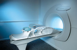
The Aurora® 1.5T Dedicated Breast MRI System with Bilateral SpiralRODEO™ is the first and only FDA approved MRI device designed specifically for breast imaging. The Aurora System, which is already in clinical use at a growing number of leading breast care centers in the US, Europe, got in December 2006 also the approval from the State Food and Drug Administration of the People's Republic of China (SFDA).
'Some of the proprietary and distinguishing features of the Aurora System include: 1) an ellipsoid magnetic shim that provides coverage of both breasts, the chest wall and bilateral axillary lymph nodes; 2) a precision gradient coil with the high linearity required for high resolution spiral reconstruction;; 3) a patient-handling table that provides patient comfort and procedural utility; 4) a fully integrated Interventional System for MRI guided biopsy and localization; and 5) the user-friendly AuroraCAD™ computer-aided image display system designed to improve the accuracy and efficiency of diagnostic interpretations.'
Device Information and Specification
CLINICAL APPLICATION
CONFIGURATION
Short bore compact
IMAGING MODES
Bi-lateral RODEO fat suppression, high resolution Rotating Delivery of Excitation Offresonance Spiral, integrated targeting SW compatible with major MR guided intervention equipment
TR
10 ms for gradient echo and less than 2,500 ms for T2 weighted spin echo
TE
From 5 ms for RODEO Plus to over 80 ms, 120 ms for T2 sequences
SINGLE/MULTI SLICE
Around 0.02 sec for a 256x256 image, 12.4 sec for a 512 x 512 x 32 multislice set
FOV
20 - 36 cm, max. elliptical 36 x 44 cm
MEASURING MATRIX
512 x 512
BORE DIAMETER
or W x H
64 cm diameter (gantry)
MAGNET WEIGHT
8,500 lbs
H*W*D
240 x 188 x 163 cm
POWER REQUIREMENTS
150A/120V-208Y/3 Phase//60 Hz/5 Wire
COOLING SYSTEM
Helium for magnet, distilled/de-ionized water for coil;
spacer

• View the NEWS results for 'Aurora® 1.5T Dedicated Breast MRI System' (3).Open this link in a new window.

• View the DATABASE results for 'Aurora® 1.5T Dedicated Breast MRI System' (2).Open this link in a new window

 
Further Reading:
  News & More:
Aurora Imaging Technology Announces Approval in China
   by salesandmarketingnetwork.com    
Aurora Imaging Technology, Inc. Release: Results of a Multi-Center Trial Demonstrates Superior Diagnostic Performance of the Aurora® 1.5T Dedicated Breast MRI System Over Whole-Body Breast MRI
Monday, 1 October 2012   by www.biospace.com    
Automatic Bolus DetectionInfoSheet: - Sequences -
Intro, Overview, 
Types of, 
etc.
 
Automatic bolus detection is used to trigger the begin of the MRI scan with the time the contrast agent reaches the region of interest. The acquisition process is started once the signal is increased in this region. There are similar methods used by the scanner manufacturer (see MRI Acronyms for Automatic Bolus Detection). After injection of the contrast agent the region of interest is monitored (e.g. with a spin echo or gradient echo sequence). When the signal increases the scan is automatically triggered or the operator is informed.

See also Abdominal Imaging, Bolus Injection, Fluoroscopic Triggering, Care Bolus, and Bolus Tracking.
spacer

• View the DATABASE results for 'Automatic Bolus Detection' (4).Open this link in a new window

 
Further Reading:
  Basics:
Fast Contrast Enhanced Imaging with Projection Reconstruction(.pdf)
   by ece.ut.ac.ir    
  News & More:
Abdominal MR angio: fast, reproducible, and safe
   by www.diagnosticimaging.com    
MRI Resources 
Hospitals - Most Wanted - Veterinary MRI - Musculoskeletal and Joint MRI - Collections - Shoulder MRI
 
previous       66 - 70 (of 73)      next 
Ab-Ab   Ab-Ac   Ac-Ac   AD-Ad   AI-Al   Al-AM   Am-Am   Am-An   An-An   An-Ap   Ap-Ar   Ar-Ar   Ar-As    At-Au    Au-Ax   
 Random Page
 
Share This Page
FacebookTwitterLinkedIn

MR-TIP    
Community   
User
Pass
Forgot your UserID/Password ?    


MRI is trending to low field magnets :
reduced costs will lead to this change 
AI will close the gap to high field 
only in remote areas 
is only temporary 
never 

Look
      Ups


MR-TIP.com uses cookies! By browsing MR-TIP.com, you agree to our use of cookies.

Magnetic Resonance - Technology Information Portal
Member of SoftWays' Medical Imaging Group - MR-TIP • Radiology-TIP • Medical-Ultrasound-Imaging • 
Copyright © 2003 - 2024 SoftWays. All rights reserved. [ 17 April 2024]
Terms of Use | Privacy Policy | Advertising
 [last update: 2024-02-26 03:41:00]